Posts

Introduction to the Electrocardiogram

Image
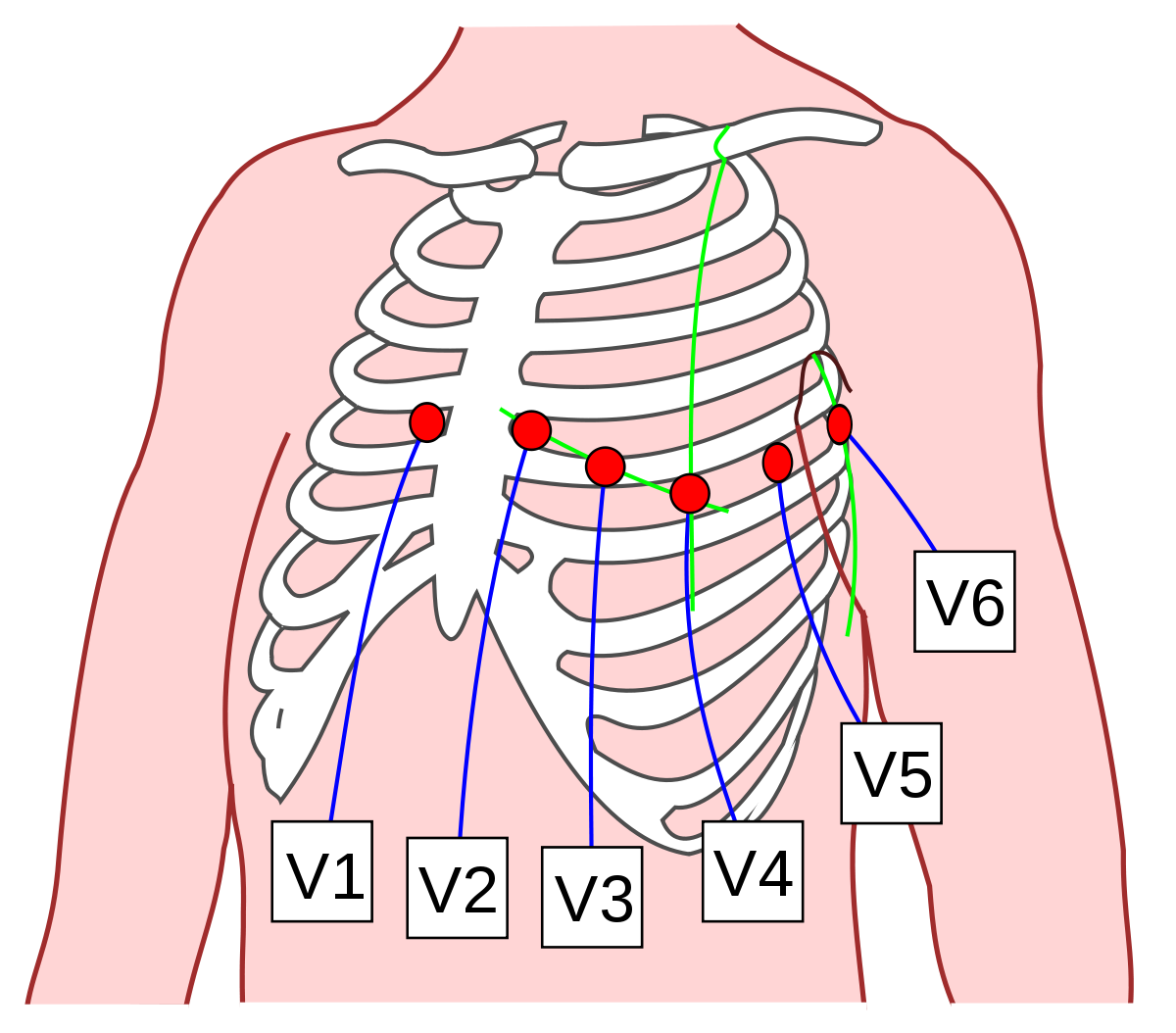
     An electrocardiogram (ECG or EKG) is a test of heart function. It measures the heart's electrical activity, indicating how well each heart chamber is contracting and ejecting blood. Recall from my previous post that the heart's autorhythmic cells can generate their own action potentials which result from ion movement across cells. Ion movement results in a difference in charge across the heart, producing a potential difference. An ECG, a graph of voltage versus time, represents the combined action potentials that autorhythmic and contractile cells generate at a particular time.       The action potential initiated by the heart's sinoatrial node spreads not only through the heart but also throughout the body. In an ECG, electrodes measure heart electrical activity from the chest, arms, and legs, enabling us to observe cardiac electrical activity from multiple locations. An essential point about ECG electrodes is that they do not produce or send electricity into the b

The Heart's Electrical Conduction Pathway

Image
     The heart is a remarkable organ. It can pump blood in an intrinsic fashion, which means that it does not depend on nerve signals to contract rhythmically. If all nerves supplying the heart are severed, the heart can still beat, as observed in patients who receive heart transplants. The heart has a conduction system that enables it to contract periodically.       The heart has two cell types: contractile and autorhythmic. Contractile cells have a stable resting membrane potential because they contract only after receiving nerve signals. They are not intrinsic and only depolarize when cells around them have depolarized. On the other hand, autorhythmic cells can contract independently of the nervous system and can depolarize by themselves. This principle is called automaticity. Unlike contractile cells, autorhythmic cells have an unstable resting potential that constantly fluctuates and reaches the depolarization threshold. These membrane potential fluctuations are called pacemak

Structure of a Red Blood Cell

Image
Red blood cells, also called erythrocytes , are fundamental to fulfilling the body's oxygen demands. They can carry up to 1.2 billion molecules of oxygen at a time! Erythrocytes are shaped like biconcave discs. This shape maximizes their surface area to volume ratio, allowing oxygen to diffuse across the red blood cell membrane efficiently.  A protein called spectrin helps erythrocytes maintain their shape and flexibility, enabling them to adjust their shape as they travel through capillaries smaller than their own diameter and to return to their original shape as they reach larger vessels.  A feature that makes erythrocytes unique is their absence of cell organelles. This maximizes the room available for oxygen to bind hemoglobin. In addition, because erythrocytes lack mitochondria, they do not utilize the oxygen they carry for their own cellular needs, making them excellent oxygen transporters. However, this also means that they cannot undergo mitosis. As they become old and rigi

COVID-19 Relief Fundraiser

Image
My brother and I have teamed up to start a food and funds initiative for Second Harvest food bank. Please visit our fundraiser page. Every dollar counts and you can make a difference, so please donate whatever you can.     Second Harvest Food Bank (The link will open in a new window) Here is a glimpse of our fundraiser.  Food and the thank you notes for our healthcare heroes.  Thank you to the children in my neighborhood who created these lovely notes. 

COVID-19 relief mask project

Image
Check out our mask sewing project. We sewed and donated over 150 masks.   

Veins and Vascular Anastomoses

Image
Arteries distribute blood to specific regions of the body. Once oxygen-rich blood reaches capillary beds, gas exchange occurs between capillaries and tissues. After gas exchange has occurred, how does carbon dioxide-rich blood return back to the heart? The answer lies in a network of vessels about 80,000 miles long: veins ! Veins tend to merge into larger vessels as they get closer to the heart. Their walls get thicker and their diameter of their lumen (central portion of blood vessels through which blood travels) also increases. Venules,  the smallest veins, have a diameter ranging from 0.008 to 1 mm. Postcapillary venules*  drain capillary beds. Just like porous capillaries, they are permeable to fluids and white blood cells. During inflammation, white blood cells often adhere to the endothelium of the postcapillary venule, and then they diffuse through the venule's wall to reach the inflamed tissue.  Figure 2: Venules drain capillary beds and gradually merge into larger veins As

Microcirculation Through Capillary Beds

Image
Capillary beds are found in nearly every tissue and organ of the body. Capillaries are minuscule exchange sites where oxygen, nutrients, and hormones pass from the bloodstream into the interstitial fluid (space between capillary walls and cells), and carbon dioxide and metabolic wastes pass from the interstitial fluid back to the bloodstream.  A  capillary bed  is an interconnected network of capillaries. Capillary beds contain different types of capillaries (read about continuous, fenestrated, and sinusoidal capillaries in my previous blog) depending on the organ in which they are located. Blood flow through a capillary bed is called  microcirculation. A capillary bed consists of true capillaries , which are the hundreds of tiny capillaries in which gas and nutrient exchange occurs between the capillary wall and interstitial fluid, and a vascular shunt , which bypasses the true capillaries.  The main arteriole that leads into the capillary bed is called the terminal arteriole . The te