An electrocardiogram (ECG or EKG) is a test of heart function. It measures the heart's electrical activity, indicating how well each heart chamber is contracting and ejecting blood. Recall from my previous post that the heart's autorhythmic cells can generate their own action potentials which result from ion movement across cells. Ion movement results in a difference in charge across the heart, producing a potential difference. An ECG, a graph of voltage versus time, represents the combined action potentials that autorhythmic and contractile cells generate at a particular time.
The action potential initiated by the heart's sinoatrial node spreads not only through the heart but also throughout the body. In an ECG, electrodes measure heart electrical activity from the chest, arms, and legs, enabling us to observe cardiac electrical activity from multiple locations. An essential point about ECG electrodes is that they do not produce or send electricity into the body; they only measure the heart's electrical activity through the chest and limbs, making the ECG safe.
Where are electrodes placed?
The conventional ECG, also called the 12-lead ECG, uses 10 electrodes and 12 leads. 1 electrode is placed on each limb and six electrodes are placed on the chest. Electrodes are designated by the letter V.
Limb electrodes can be inserted anywhere on the limbs as long as they are symmetrical and are not placed over bone.
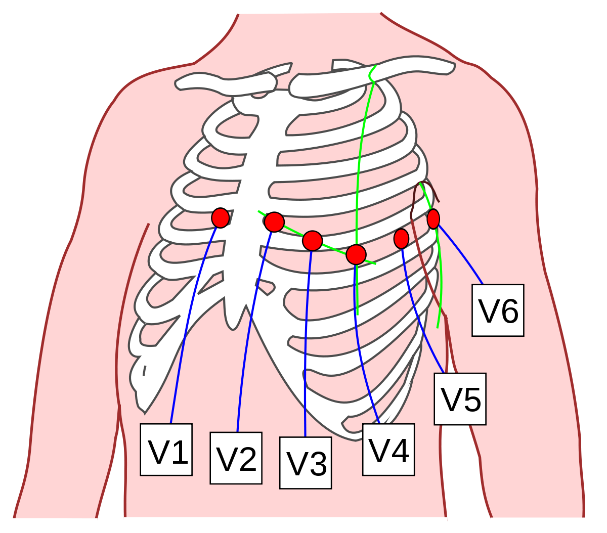
The chest electrodes, also called precordial electrodes, are placed in the space between two ribs, called the intercostal space.
- The first and second electrodes, V1 and V2, are placed in the fourth intercostal space on both sides of the sternum.
- The third electrode, V3, is placed between V2 and V4.
- The fourth electrode, V4, is placed in the fifth intercostal space on the midclavicular line.
- The fifth electrode, V5, is placed in the fifth intercostal space on the anterior axillary line.
- The sixth electrode, V6, is placed in the fifth intercostal space on the mid-axillary line.
Source: Wikimedia Commons
A
lead is a graphical representation of the heart's electrical activity obtained from multiple electrodes. It measures the difference in electric potential between two different regions. A bipolar lead measures the potential difference between a positive and negative electrode. A unipolar lead measures the potential difference between a positive electrode and a reference point (that is not negative). An ECG consists of six limb leads and six chest leads. The Bipolar Limb Leads
In the Lead I configuration, a positive electrode is placed on the left arm and a negative electrode is placed on the right arm. Lead I looks at cardiac electrical activity from the left.
In the Lead II configuration, a positive electrode is placed on the left leg and a negative electrode is placed on the right arm. Lead II looks at cardiac electrical activity from the inferior left perspective.
In the Lead III configuration, a positive electrode is placed on the left leg and a negative electrode is placed on the left arm. Lead III looks at cardiac electrical activity from the inferior right perspective.
The Unipolar Limb Leads
The unipolar limb leads - also called the augmented limb leads - use one limb electrode as the positive electrode. The midpoint between the remaining two limb electrodes is used as the reference point. The three unipolar limb leads are the augmented vector right (aVR), augmented vector left (aVL), and augmented vector foot (aVF) leads.
aVR lead measures the potential difference between a positive electrode on the right arm and a reference point midway between the left arm electrode and the left leg electrode.
aVL lead measures the potential difference between a positive electrode on the left arm and a reference point midway between the right arm and left leg.
aVF lead measures the potential difference between a positive electrode on the left leg and a reference point midway between the right arm and left arm.
The Unipolar Chest (Precordial) Leads
The precordial leads use the six precordial electrodes (V1-V6) as the positive electrodes and the midpoint of the three limb leads as the reference point.
References:
https://www.ecgedu.com/proper-electrocardiogram-ecg-ekg-lead-placement/
https://youtu.be/kwLbSx9BNbU


Comments
Post a Comment